 呼吸器 13. サルの肺の概観--
サル、フォルマリンの還流固定と気管系の注入固定、セロイディン包埋、
呼吸器 13. サルの肺の概観--
サル、フォルマリンの還流固定と気管系の注入固定、セロイディン包埋、
H-E染色、x8.------------------------------------------------
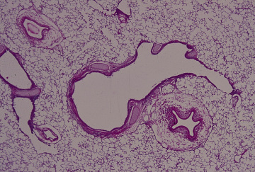
これはサルの一つの肺葉の中心部で、画面の中央部を占める大きな管が-
気管支枝、これは右方に伸びて三本の終末細気管支に分かれる。その右-
端の管では壁が所々で消えて、肺胞になっている。この部分が呼吸細気--
管支である。この気管支枝の壁には軟骨と平滑筋が認められる。気管支--
枝の右下に接しているのは肺動脈の枝。画面の左端には細い終末細気管-
支とこれに続く呼吸細気管支が認められる。画面の左上の独立した血管--
は肺静脈の枝である。画面の残りの部分は肺胞で満たされている。------
Respiratory system 13. Lung
of a monkey. General view.-----
Monkey, perfusion and infusion
fixation with formalin, embedding with -------
celloidin, H-E stain, x 8.---------------------------------------------
This is the central portion of a macaque pulmonary lobe. At center
of --------
the field, there is a branch of bronchiole, which extends right
and divides ------
into 3 terminal bronchioles. Among these the rightmost one continues
into ----
a respiratory bronchiole. Lower right to this branch of bronchiole
there is -----
a branch of pulmonary artery. At leftmost of the field one terminal
bronchiole -
and its continuation, respiratory bronchiole, are seen. At upper
left a branch --
of pulmonary vein is seen. Except for these structures, whole
field is occupied -
with alveoli. --------------------------------------------------------
戻る