 呼吸器 15. 呼吸細気管支と肺胞管.
サル、フォルマリンの還流固定と気管系の注入固定、セロイディン包埋、
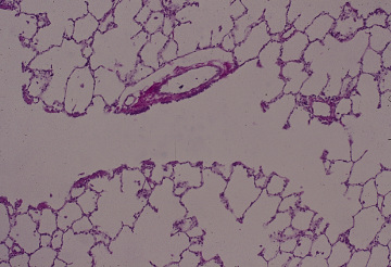
呼吸器 15. 呼吸細気管支と肺胞管.
サル、フォルマリンの還流固定と気管系の注入固定、セロイディン包埋、
H-E染色、x50.-----------------------------------------------
これは「呼吸細気管支1」の右半分の拡大である。画面の中央部に位置し、
上方と下方に、壁が途切れ途切れに見られる部分が呼吸細気管支である。
これはその右端で、右下に伸びる管となるが、この管の壁はすべて肺胞--
でできている。これが肺胞管である。--------------------------------
Respiratory system 15. Respiratory
bronchiole and alveolar duct..
Monkey, perfusion and infusion
fixation with formalin, embedding with
celloidin, H-E stain, x 50.-------------------------------------
This is the higher magnification of right half of Respiratory
bronchiole 1.------
In the middle of this field one respiratory bronchiole lies horizontally
from ---
left to right. The left half of it, having the wall at intervals
in both upper and -
lower side, is the respiratory bronchiole which terminates as
an alveolar duct
at lower right corner of the field. -------------------------------------
戻る