 呼吸器 20. 呼吸細気管支と肺胞管
ヒト、フォルマリン注入固定、ハラフィン包埋、H-E染色、x
40.
呼吸器 20. 呼吸細気管支と肺胞管
ヒト、フォルマリン注入固定、ハラフィン包埋、H-E染色、x
40.
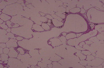
画面の中央を右上から左下に走る管が肺胞管である。この管の右上端の
円形の部分は繊毛上皮で被われており、終末細気管支である。この円形-
の管の左端の部分で、その上皮が消えて肺胞が始まり、呼吸性細気管支
となる。更に左下方に進むと肺胞だけで囲まれた管、即ち、肺胞管とな---
る。ヒトの肺は巨大であるので、このような気管支樹の構造を連続的に---
観察できる機会は稀である。--------------------------------------
Respiratory system 20. Respiratory
bronchiole and alveolar duct.
Human, infusion fixation with
formalin, embedding with paraffin,
H-E stain, x 40.---------------------------------------
In this figure one respiratory channel from terminal bronchiole
to the alveolar --
duct is continuously observed. At upper right is a terminal bronchiole
being lined
with ciliated columnar epithelium intermingled with goblet cells
and at lower left--
portion this bronchiole opens into a respiratory bronchiole which,
in turn, into an-
alveolar duct whose wall consists exclusively of alveoli. Because
of huge size of -
human lung such a case is seldom encountered to be able to follow
a respiratory
channel continuously. ---------------------------------------------
戻る