 Œؤ‹zٹي 22. ”x–Eٹا
ƒqƒgپAƒtƒHƒ‹ƒ}ƒٹƒ“’چ“üŒإ’èپAƒnƒ‰ƒtƒBƒ“•ï–„پAH-EگُگFپA‚ک
100.
Œؤ‹zٹي 22. ”x–Eٹا
ƒqƒgپAƒtƒHƒ‹ƒ}ƒٹƒ“’چ“üŒإ’èپAƒnƒ‰ƒtƒBƒ“•ï–„پAH-EگُگFپA‚ک
100.
گ}‚ج‰Eڈم’[•”‚©‚çچ¶‰؛•û‚ةگL‚ر‚éٹا‚ھ”x–Eٹا‚إ‚ ‚éپB”x–Eٹا‚ج•ا‚ئ‚µ‚ؤ‚حپA
ŒآپX‚ج”x–E‚ھ‹¤’ت‚جٹاچo‚ةٹJ‚ٹJŒû•”‚ًژو‚èٹھ‚¢‚ؤ‚¢‚éپAڈ—ت‚ج•½ٹٹ‹ط‘@--
ˆغ‚ھ”F‚ك‚ç‚ê‚é‚ج‚ف‚إ‚ ‚éپB”x–Eٹا‚ح‚»‚ج‰“ˆت’[‚إگ”–{‚ة•ھ‚©‚êپA‹¤’تچo--
‚ً”x–E‚¾‚¯‚ھˆح‚قگو‚ھچs‚«ژ~‚ـ‚è‚ج‚س‚‚ëپA”x–E”X‚ئ‚ب‚ء‚ؤڈI‚ي‚éپB”x–E----
”X‚إ‚ح”x–E‚جٹJŒû•”‚ة•½ٹٹ‹ط‘@ˆغ‚حŒ©‚ç‚ê‚ب‚¢پB----------------------
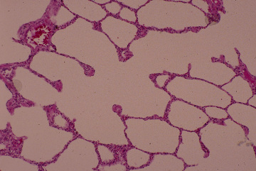
Respiratory system 22. Alveolar
duct.
Human, infusion fixation with
formalin, embedding with paraffin,
H-E stain, x 100.---------------------------------------
In this figure an alveolar duct runs from upper right to lower
left. As the wall of -
alveolar ducts we can only observe the smooth muscle fiber bundle
encircling the
opening of each alveolus into the common lumen of alveolar duct.
The alveolar ---
duct divides into several number of alveolar sacs, consisting
exclusively of alveoli.
In alveolar sac, there are no encircling smooth muscle fibers
at alveolar opening.
–ك‚é