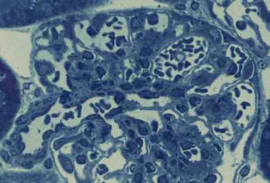
腎臓 18. 腎小体(エポン切片)
ラット、グルタールアルデヒド・パラフォルムアルデヒドで還流固定、エポン包埋、
トルイディンブルー染色、x 750.---------------------------------------------------------------------
これは還流固定したラットの腎臓の厚さ1μmの切片である。標本が薄いので、細胞
の重なりがなく、個々の細胞を明瞭に識別できる。画面の左下から右上方に輸入細動
脈が進入する。輸入細動脈では内皮細胞の核と平滑筋細胞の核が識別できる。輸入細
脈は毛細血管となって糸球体を構築するが、毛細血管の中に少数の赤血球が残ってい
るので、毛細血管の内腔とこれを縁取る内皮細胞の核が明らかに認められ、毛細血管
の外に付着してボウマン腔に突出している「足細胞」の識別も容易である。また、特
に図の上半部では毛細血管を束ねる結合組織の中に、輪郭に小さい凹凸を示すメザン
ギウム細胞の核が見られる。生体を還流固定して作った標本では、糸球体とボウマン
嚢の間の隙間は非常にせまい。――――――――――――――――――――――――
Kidney
18. Renal corpuscle (Epon section).
Rat, perfusion fixation with Glutaraldehide-Paraformaldehide fixative, embedding--
with
Epon, toluidinblue stain, x 750.^^^^^^^^^^^^^^^^^^^^^^^^^^^^^^^^^^^
As
this is a very thin section(ca. 1μm), fine structures are clearly seen. At lower left―
the afferent
arteriole goes into the renal corpuscle and forks abruptly into capillaries―
to-form the glomerulus. Capillaries contain some
erythrocytes so that the capillary――
lumen
and nuclei of endotheliar cells are easily recognized. The podocytes attaching----
outside to the capillaries and having a large nucleus and densely stained cytoplasm are
also perceived without any difficulties. In the upper half of this field some nuclei of the
mesangium
cells, showing irregular contour, are noted in the connective tissue that----
bundles
the capillaries. In the specimens fixed with perfusion, the space between-------
Bowman’s
capsule and glomerulus is very narrow.-----------------------------------------------