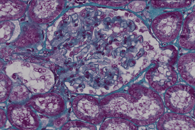
腎臓 7. 腎小体及び尿細管 1.
ヒト、ブアン液で固定、パラフィン包埋、M-G染色、x 250.
これは急速な出血によって死亡した遺体から得られた標本で、細胞及び組織の保存が
非常に良い。また厚さ約3μmの薄い標本であるので、腎小体と尿細管の微細構造を
よく理解できる。画面の中央に1個の腎小体があり、その周囲を尿細管、その殆どが-
曲尿細管の主部(近位曲尿細管)、が埋めている。腎小体は毛細血管の集合体である糸
球体と、その周囲を包む糸球体嚢(ボウマン嚢)からなり、一方の極(血管極、図の
左側)から輸入細動脈と輸出細動脈が出入し、反対側の極(尿細管極、図の右側)か
ら尿細管が始まる。輸入細動脈は血管極において数本の毛細血管に分かれるが、これ
らは枝分かれも互いの吻合もすることなく、ループ状に走って血管極に帰り、1本の
輸出細動脈にまとまって腎小体を去る。この毛細血管のループ全体をまとめて糸球体
という。輸入細動脈と輸出細動脈とが作るV字型のくぼみに(左から)接している管
が、尿細管の介在部のうちの特に緻密斑と呼ばれる部分である。糸球体の毛細血管の
表面に密着してこれを被う被蓋細胞(「足細胞、podocytes」ともいう)は、血管極に
おいて反転して糸体球嚢の内面を被う単層扁平上皮となる。この単層扁平上皮は尿管
極において突然単層立方上皮である尿細管主部の上皮に移行する。「腎臓 8,9,10」は
この腎小体の拡大像、また「腎臓11〜13」も同じ切片の写真である。―――――――
Kidney
7. Renal corpuscle and urinary tubules 1.
Human, fixation with Bouin’s fluid, embedding with paraffin, M-G stain, x 250.
Since
this specimen was obtained from a female, who died by rapid bleeding, cells and----
tissues
are very well preserved, and because of thinness of this section of about 3μm-----
fine
structures of kidney are clearly seen. In the middle of this field there is a
renal--------
corpuscle
and surrounding space of it is filled with urinary tubules, mainly proximal-------
convolutions.
A renal corpuscle consists of a glomerulus and a Bowman’s capsule. At-------
one (vascular) pole un afferent arteriole goes into the corpuscle and un efferent arteriole
goes
out. At the opposite (urinary) pole un urinary tubule begins. At the vascular
pole-----
the
afferent arteriole suddenly divides into several numbers of capillaries, each
of that----
runs
in loop and unites with each other again at the vascular pole to form the
efferent----
arteriole.
The whole of these capillaries is named as glomerulus. The inner surface of------
the
Bowman’s capsule is covered with simple squamous epithelium, which changes at-----
the
vascular pole into the specified covering cells of capillaries, i.e. podocytes,
and at the---
urinary
pole suddenly changes into the simple cuboidal epitheliar cells of the urinary-------
tubule. The narrow space between glomerulus and Bowman’s capsule is called---------------
Bowman’s
space. “Kidney 8,9,10.” are higher magnifications of this renal corpuscle and----
“Kidney
11~13” are also taken from this preparation.-------------------------------------------------