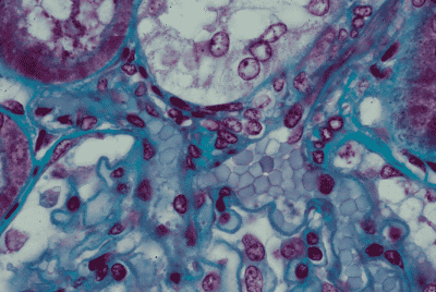
腎臓 8. 腎小体の血管極 1.
ヒト、ブアン液で固定、パラフィン包埋、M-G染色、x 600.
これは「腎臓 7.」の血管極の拡大である。画面の右上から左下方に下りてくるのが
輸入細動脈で、その流入部の左に接してほぼ真上に向かって走り、腎小体を出ると
すぐ左に曲がっているのが輸出細動脈である。輸入細動脈の壁には平滑筋繊維が認
められるが、輸出細動脈の壁には認められない。輸入細動脈は腎小体に入ると直ち
に数本の毛細血管に分かれ、これらはループ状に走って血管極の近くで再び合一し
て輸入細動脈となる。画面下部の中央やや右に3個の大きな明るい核が見られるが、
これが「足細胞」の核である。糸球体嚢の内面を被う扁平上皮細胞の核は、このボ
ウマン嚢の右上部と左上部に各1個認められる。ボウマン嚢の繊維皮膜が血管極で
反転して糸球体の表面に移行している像も明瞭である。輸入細動脈と輸出細動脈が
挟むくぼみに接して、明るい胞体と4個の円形の核が認められるが、これが介在部
の緻密斑である。これと輸入・輸出細動脈の間には数個の核が密に存在し、特に輸
入細動脈に近い右側ものは無色透明な胞体の中に円形の核を持つが、これらは傍糸
球体細胞と呼ばれ、血圧上昇作用を持つレニンを分泌すると考えられている。-------
Kidney
8. Vascular pole of a renal corpuscle 1.
Human, fixation with Bouin’s fluid, embedding with paraffin, M-G stain, x 600.
This is a high power magnification of “Kidney 7.” From upper right to lower left comes
an
afferent arteriole and divides into several numbers of capillaries, that in
turn unite--―
together
to form an efferent arteriole which goes out at the top of this renal corpuscle-----
and
soon turns to left. In the wall of the afferent arteriole some smooth muscle
fibers------
are discernible but in the wall of the efferent arteriole not. At lower middle portion of------
this field 3 large nuclei attach to the capillary surface, that are nuclei of podocytes. Two----
flattened
nuclei locating each left and right at upper portion of the Bowman’s capsule------
are
that of covering epitheliar cells. In the V shaped hollow between afferent and-----------
efferent
arterioles there are 4 round nuclei in the pale cytoplasm are seen; they are--------
constituting
cells of macula densa. Between macula densa and afferent and efferent--------
arterioles
several numbers of flattend nuclei and cells with round nuclei and pale------------
cytoplasm
are recognizable. They are called as a whole juxta-glomerular cells and-----------
believed
to segregate the hormone, Renin.------------------------------------------------------------