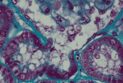
腎臓 9. 腎小体の尿管極 1.
ヒト、ブアン液で固定、パラフィン包埋、M-G染色、x 600.
これは「腎臓 7.」の尿管極の強拡大である。画面の上部中央に糸球体の下端部が見え、-----
その右上部に、毛細血管の表面に付着している2個の「足細胞」が認められる。この--------
糸球体の左右を囲むようにボウマン嚢の繊維皮膜とその内面を被う扁平上皮が認められ、---
この上皮細胞は尿管極で突然、尿細管主部の赤色に濃染する単層立方上皮に移行する。------
主部の上皮は丈が高く、自由表面に著明な刷子縁をそなえており、管腔は比較的狭い。------
画面の右下に典型的な主部の横断面が1個見られる。ただし、この標本では上皮の丈は-----
比較的低く、管腔も広い。尿細管の周囲は多数の毛細血管で埋められている。-----------------
Kidney
9. Urinary pole of a renal corpuscle 1.
Human, fixation with Bouin’s fluid, embedding with paraffin, M-G stain,x 600.
This
is a high power magnification of “Kidney7”. The upper middle portion of this---------
field
is occupied by the lowermost portion of the glomerulus. At upper right 2
podocytes--
attaching
to the capillary surface are seen. At right and left facing to the glomerulus------
across
the Bowman’s space, Bowman’s capsule consisting of fibrous capsule and inner―-
covering
epitheliar cells is noted. The inner covering cells change suddenly at the----------
urinary
pole into the epitheliar cells of the beginning portion of the urinary tubule,--------
proximal
convolutions. These cells are simple cuboidal in shape, deeply stained with-------
acid
dye and provide with conspicuous brush border at free surface but no cell
boundary-
among
them is discernible. At lower right corner there is a typical transverse
section of---
the
proximal convolutions. Urinary tubules are surrounded by so many capillaries.--------