 肝臓 16.格子繊維 1.
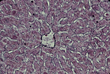
サル、フォルマリン固定、パラフィン包埋、鈴木鍍銀法、
ケルンエヒトロートで後染色、x 64.----------------
肝臓の実質細胞である肝細胞は連なって索を作り、これが左右にも----
上下にも複雑に連結してスポンジ状の網工を形成する。--------------
この網工の隙間は、管腔の広い特殊な毛細血管である類洞(洞様血管)
で埋め尽くされている。肝細胞索と類洞の間にはごく少量の結合組織---
繊維が介在する。この繊維は銀に対して強い親和性を持ち、各種の----
錯銀液で処理すると黒染する。格子繊維を染めると、類洞の輪郭が----
明瞭になる。肝臓ではこの繊維の配列のパターンが格子状であるので、
格子繊維と呼ばれる。図の中心には中心静脈の横断面があり、これに-
流入する類洞の配列が明瞭である。肝臓9. はこれの強拡大である。---
Liver 16. Reticular fibers 1.
Monkey, fixation with formalin, embedding with paraffin, silver --
impregnation after Suzuki, counter stain with Kernechtrot, x 64.
Hepatic cell cords anastomose with neighboring ones so that ----
they constitute complex 3-dimensional meshworks, which--------
intermingled with that of special capillary vessels, sinusoids.------
Between hepatic cell cords and sinusoids lie a few reticular ------
fibers, that have intense affinity with silver ion. In this figure
-----
reticular fibers are demonstrated by a silver impregnation -------
method so that the contour of sinusoids is evidently recognizable.
戻る