 肝臓 17. 格子繊維 2.
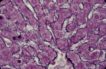
サル、フォルマリン固定、パラフィン包埋、鈴木鍍銀法、
ケルンエヒトロートで後染色、x 160.---------------
これは肝臓6.の拡大である。下辺中央部にある中心静脈とこれに---
注ぐ洞様血管(類洞)、ならびに類洞を取り巻く格子繊維(銀好性繊維)
が明瞭に観察される。また類洞と実質細胞である肝細胞索との密接な
関係も明らかである。----------------------------------------
Liver 17. Reticular fibers 2.
Monkey, fixation with formalin, embedding with paraffin, silver-impregnation
after Suzuki, counter-stain with Kernechtrot, x 160.-------------------
This is higher magnification of Fig. Liver 6. At lower middle
portion is -----
the central vein, into which several sinusoids flow. Reticular(argyrophilic)
--
fibers enveloping sinusoids are clearly demonstrated and intimate
--------
relationship between sinusoids and hepatic cell cords is also
conspicuous. --
戻る