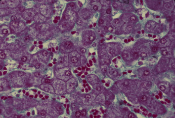
 肝臓 18. 肝細胞索と類洞.
ヒト、ブアン液で固定、パラフィン包埋、マッソン-ゴールドナー(M-G)染色、x 160.
M-G染色では細網繊維を青緑色に,肝細胞を赤く、赤血球を赤橙色に----
染め分けるので、肝細胞索と類洞とを明瞭に識別できる。この図では、---
類洞の中に赤橙色に染まった赤血球が適当に含まれているので、類洞が-
特に明らかである。肝細胞索では、胆毛細管がここかしこで観察される。--
Liver 18. Hepatic cell cords
and sinusoids.
Human, fixation with Bouin's fluid, embedding with paraffin, M-G
stain, x 160.
As M-G stain stains reticular fibers bluish green, hepatic cells------
red and erythrocytes reddish orange, we can easily distinguish------
hepatic cell cords from sinusoids. As in this preparation sinusoids----
contain properly erythrocytes, sinusoids are especially clear.
-------
Among the hepatic cell cords bile canaliculi are seen here and
there. -
戻る