 肝臓 24.ヘリング管3.縦断面.
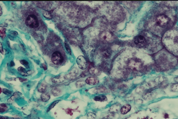
ヒト、ブアン液で固定、パラフィン包埋、M-G染色、x 400.
これはヘリング管の縦断面である。画面の中央から左下方に伸びる、---
ほとんど無色の管がヘリング管で、2列に並ぶ無色の上皮細胞の間に、--
その管腔が明瞭に認められる。この管腔の右上端部に上方の肝細胞---
索の胆毛細管が連なっている。画面の右下を斜めに走っている無色----
の上皮細胞からなる管も、ヘリング管である。緑色に染まっている------
のは結合組織繊維。-------------------------------------------
Liver 24. Canal of Hering 3.
Longitudinal section.
Human, fixation with Bouin's fluid, embedding with paraffin, M-G
stain, x 400.
In this figure 2 canals of Hering are seen. The left one, running
from ---
the center to lower left, is quite tongitudinally cut and its
narrow lumen
is distinctly observed. At upper right end of this lumen a distinct
bile ---
canaliculus in the hepatic cell cord opens. Another canal of Hering
-----
lies at lower right corner of this figure. ----------------------------
戻る