 肝臓 28. 類洞
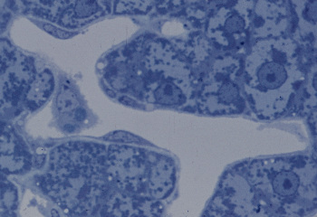
サル、グルタルアルデヒドで還流固定、エポン切片、トルイディンブルー染色、x 400.
類洞は肝小葉の中で肝細胞索と複雑に絡み合う特殊な毛細血管で、-----
内腔は広いが、それを縁取る壁は内皮細胞のみである。この標本は-----
還流固定したサルの肝臓をエポキシ樹脂に包埋して、厚さ約1ミクロン----
の薄い切片にしたもので、類洞を縁取る極めて薄い内皮細胞の細胞体と、-
これに開いている小孔、および内皮細胞と肝細胞の間の狭い隙間(これを-
ディッセ腔という)が明らかに認められる。類洞の中には1個の星細胞-----
が認められる。肝細胞では核の周囲に明るい領域が見られるが、これは--
グリコーゲンが密集している領域である。----------------------------
Liver 28. The sinusoid.
Monkey, perfusion with glutaraldehyde-fixative, embedding with
epon,
toluidine blue stain, x 400. ------------------------------------
Because of perfusion fixation and thinness of the section, the
fine---
structures of sinusoids and hepatic cells are well recognizable.
-----
Very thin cytoplasm of endotheliar cells, minute openings therein
---
and very narrow space between sinusoids and hepatic cells, space
--
of Disse, are clearly seen. In the sinusoid a Kupffer's stellate
cell ---
is recognized.----------------------------------------------
戻る