 肝臓4.肝小葉の概観2.
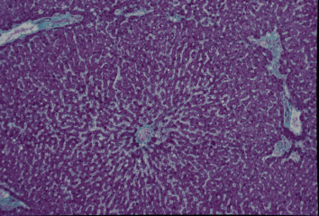
ヒト、ブアン液で固定、パラフィン包埋、マッソン-ゴールドナー(M-G)染色、x 25.
ヒトでは小葉間結合組織が少量であるために、肝小葉の輪郭が分かり難い。
この図は1個の肝小葉のほぼ全景を示している。中央やや下部に中心静脈--
があり、ここから放射状に肝細胞索が四方に広がっている。図の左上、------
左下、および右側に小葉間結合組織があり、この肝小葉を境しているが、----
下方及び上方中央部ではこの肝小葉は隣のものと連続している。-----------
Liver 4. General view of a hepatic
lobule 2.
Human, fixation with Bouin' fluid, embedding with paraffin, M-G
stain, x 25.
In human, because of scantiness of interlobular connective tissue,
-
it is not easy to recognize the contour of each hepatic lobule.-----
In this figure one hepatic lobule is shown, demarcated with--------
interlobular connective tissue at upper left, lower left and right
side.
At the upper central and lower portions this lobule continues
with --
neighboring lobules. At the center of this figure locates the
central -
vein, from which hepatic cell cords run to periphery radially.-------
戻る