 肝臓 5. 肝小葉概観 3.
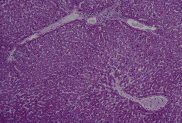
ヒト、ブアン液で固定、パラフィン包埋、H-E染色、x 25.
ヒトでは小葉間結合組織が少量であるために、肝小葉の輪郭は、---
一般に明瞭でない。この図では、左上と右上の2辺は小葉間静脈を-
含む小葉間結合組織によって比較的明瞭に境されているが、左下---
および右側では隣の小葉との間には境は認められない。図の右下の-
大きな管は小葉下静脈で、これに左上方から中心静脈が注いでいる。
この中心静脈に注ぐ洞様血管も明らかに認められる。-------------
Liver 5. General view of a hepatic
lobule 3.
Human, fixation with Bouin's fluid, embedding with paraffin, H-E
stain, x 25.
In human, since interlobular connective tissue is scanty, lobular
--
configuration is less evident. In this figure upper left as well
as ---
upper right sides are relatively well limited with interlobular------
connective tissue containing each an interlobular vein, but lower--
left side boundary of this lobule is not indicated. At the lower ----
right portion there is a sublobular vein and a central vein opening-
into the former.------------------------------------------
---------------
戻る