 肝臓 6. 小葉間結合組織 1.
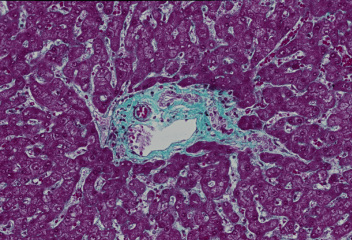
ヒト、ブアン液で固定、パラフィン包埋、マッソン-ゴールドナー(M-G)染色、x 64.
M-G染色では、結合組織が青緑色に染まり、肝細胞索が赤く染まるので、
小葉間結合組織を明瞭に観察できる。青緑色の小葉間結合組織の中に、
壁が薄くて腔が広い小葉間静脈と、円形の断面で中に赤橙色に染まった-
赤血球の詰まった小葉間動脈が明らかに識別される。類洞を取り巻く細網
繊維も青緑色に染まっているので、類洞と肝細胞索の関係もよくわかる。-
Liver 6. Interlobular connective
tissue 1.
Human, fixation with Bouin's fluid, embedding with paraffin, M-G
stain, x 64.
As the M-G stain stains connective tissue bluish green, the interlobular----
connective tissue stands out from the reddish stained hepatic
parenchyma.
In this interlobular connective tissue one interlobular vein having
a wide---
lumen and one interlobular artery containing reddish stained erythrocytes-
are clearly noted. Reticular fibers surrounding sinusoids stain
green so that
the relationship between sinusoids and hepatic cell cords is also
evident.---
戻る