 肝臓 7. 小葉間結合組織 2.------
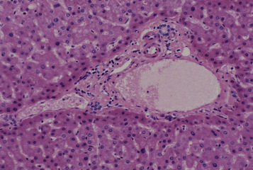
ヒト、ブアン液で固定、パラフィン包埋、H-E染色、x 66.
3個の肝小葉の会合部の小葉間結合組織間が示されている。---------------
小葉間結合組織間の中を門脈の枝である小葉間静脈と肝動脈の枝である-----
小葉間動脈と肝臓の分泌物である胆汁を運び出す小葉間胆管が通っている。--
この3種類の管をまとめて門脈の三つ組という。図の中央やや右の大きな-----
断面が小葉間静脈、左下方に伸びているのはその枝である。大きな小葉間----
静脈の上にある小さい2個の濃い桃色の管が小葉間動脈であり、その右上----
の青色に染まった核で囲まれた管が小葉間胆管である。なお壁が明瞭で------
ない裂け目のような空間はリンパ管である。小葉間結合組織に接する肝-------
小葉の辺縁部の肝細胞は他の肝細胞よりやや小型で、やや濃く染まる。-------
Liver 7. Interlobular connective
tissue 2.
Human, fixation with Bouin's fluid, embedding with paraffin, H-E
stain, x 66.
This is the interlobular connective tissue, locating at the meeting---
place of 3 lobules.In this interlobular connective tissue a large
-----
interlobular vein, two small interlobular arteries and a interlobular --
bile duct are seen. A split-like space at upper left of the interlobular
vein is a lymphatic. The hepatic cells directly surrounding this
-----
interlobular connective tissue are smaller in size and more deeply--
stained than other hepatic cells. -----------------------------
戻る