卵巣2.皮質概観.
サル、SUSA液で固定、セロイディン包埋、H-E染色、x 40.
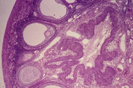
「卵巣1」の拡大。画面の左端のやや赤く見える部分が白膜で、その右に幼弱な
卵胞が密集した層が続き、更にその右には胞状卵胞が並んだ層が連なる。皮質
の深部、画面の右半分を占めているのが黄体である。
Ovary
2. General view of the ortex.
Monkey, fixation with SUSA-fluid, embedding with celloidin, H-E stain, x 40.
Higher magnification of “Ovary 1”. The somewhat reddish stained thin layer at the left
end of the field is the tunica albuginea, right to that follows the thick cortex,consisting
of numerous young egg-follicles embedded
in the cell-rich connective tissue, and then ---
developed
vesicular follicles. The right half of the field is occupied by a well
developed ---
corpus luteum.---------------------------------------------------------------------------------------------