精巣 1. 矢状断全景.
ヒト、ブアン液で固定、セロイディン包埋、H-E染色、x 3.
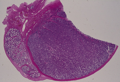
これはヒトの精巣の矢状断の全景で、画面の左が上、画面の下が前である。精巣は精子を
作り出す器官で、その形は左右にやや扁平な卵型、大きさは拇指頭大である。精巣の上部
から後面にかけて精巣上体が密着する。図の中央の大部分を占める濃い紫色の部分が精巣
で、その左に付いている紫色のやや薄い部分が精巣上体である。精巣の全表面を包む濃い
赤に染まった部分は、緻密な結合組織の皮膜(白膜)である。白膜は精巣の後上部で厚い
結合組織性の障壁(精巣縦隔)を精巣の実質内に突出させ、これは更に多数の結合組織の
索ないし薄板を前面及び側面の白膜に向かって伸ばして、精巣の内部を不完全ながら------
200~300の円錐形の区画(精巣小葉)に分けると共に、内部を支える骨組みを作る。-----
図の左上部、精巣の後上部に密着する赤い部分が精巣縦隔である。----------------------------
Testis
1. Sagittal section.
Human, fixation with Bouin’s fluid, embedding with celloidin, H-E stain, x 3.
This figure shows a whole sagittal section of a human testis. The big central portion,--―---
stained deeply violet, is testis itself, the sperm producing organ. The testis is enclosed in---
a thick fibrous capsule, tunica albuginea, which thickens on the posterior aspect of—―----
testis and projects into the organ as mediastinum testis. Numerous fibrous thin septa,---―
septula testis, extend radially from the mediastinum to the tunica albuginea dividing------
the
organ into 200~300 conical lobules, lobuli testis. Upper left the mediastinum
is seen---
as a deeply red stained dense connective tissue mass. On the left side the epididymis-------
attaches to the testis.-----------------------------------------------------------------------------------------