精巣11. 精子形成3.
サル、ブアン液で固定、パラフィン包埋、H-E染色、x 750.
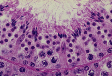
ここに見られるのは精祖細胞と精母細胞と精子細胞及びこの精子細胞の前の世代の
精子細胞が形態変化を遂げた精子である。この画面ではセルトリー細胞が特に明瞭
に観察される。即ち、画面の中央の1個から右方に2個、左方に2個のセルトリー
細胞が立ち並び、特に中央のものと左端のものでは、細胞質と、基底膜から離れて
高いところに位置する核とが明瞭である。精子細胞は成立したばかりであり、核は
未だ完全に休止期に入っていない。精母細胞はこの精子細胞の次の世代のものであ
る。基底膜に付着する比較的小型の円形の核が精粗細胞の核である。セルトリー細
胞の頂上に付着して行われてきた精子組織形成は終わりに近づき、核質が凝縮した
精子の頭とそれから上方に伸びる尾が明瞭になっている。-------------------------------
Testis
11. Spermatogenesis 3.
Monkey, fixation with Bouin’s fluid, embedding with paraffin, x 750.
Here 4 kinds of cells are recognized from the uppermost layer to the bottom, i.e.
the spermatids performing the spermatohistogenesis, the spermatids of the next
generation just formed, the primary spermatocytes of the after next generation,
and the spermatogonia. Five Sertoli cells are also clearly seen, standing on the--
basement membrane and protruding into the lumen. Their nuclei locate high---
from the basement membrane in the intensely pink stained cytoplasm.-----------