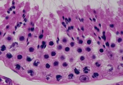
精巣15.精子組織形成4.
サル、ブアン液で固定、パラフィン包埋、H-E染色、x 750.
精子組織形成が進み、核は濃縮されて小さくなり、精子の頭部の形に近づき、
セルトリー細胞の細胞質に付着している。精子細胞の細胞質は反対に管腔側に
偏在する。精子の頭部から生じた尾がこの細胞質を貫いて管腔の中に伸びる。
「精子組織形成3」で見られた次の世代の精母細胞が分裂して精娘細胞に替わ---
っている。基底膜に接する最下層では精祖細胞の分裂像が見られる。------------
Testis
15. Spermatohistogenesis 4.
Monkey, fixation with Bouin’s fluid, embedding with paraffin, x 750.
The spermatohistogenesis progresses further and nuclei of spermatids become-------
smaller in size and spindle in shape approaching to the sperm-heads.--------------------
The sperm-tail appears which penetrate the cytoplasm projecting into the lumen.----
The primary spermatocytes, seen beneath the spermatids in “Spermatohistogenesis-
3”, performed the first meiotic division and appear the secondary spermatocytes. In-
the lowermost layer, attaching the basement membrane mitotic figures of the--------
spermatogonia are seen.----------------------------------------------------------------------------