精巣16.精子組織形成5.
サル、ブアン液で固定、パラフィン包埋、H-E染色、x 750.
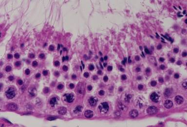
精子組織形成が終わりに近づいた。精子細胞の核は濃縮して精子の頭部となり、
セルトリー細胞に付着している尖端の反対側から出発した尾は長く伸びて管腔----
に突出する。不要になった細胞質は精子から離断して、断片となっている。精----
子の頭部の下層には成熟分裂が終わったばかりの、次の世代の精子細胞が3~4----
列並び、さらにその下層には2世代次の精母細胞が認められる。基底膜に接し----
ている円形の核は精粗細胞の核である。画面の中央のやや左に、核と細胞質が-----
明瞭なセルトリー細胞が認められる。この図に続く次の段階が「精巣11」に示---
されている。------------------------------------------------------------------------------------
Testis
16.Spermatohistogenesis 5.
Monkey, fixation with Bouin’s fluid, embedding with paraffin, x 750.
This
is the end-stage of spermatohistogenesis. The nucleus of the spermatids-----------
has been highly concentrated and become sperm-head. Attaching to the cytoplasm-----
of
Sertoli cells at the head top, sperms perform further maturation: the tails get--------
longer and protrude highly into the lumen of the seniniferous tubule and cytoplasm---
of spermatids no longer necessary to sperm is separated from sperms to become -------
debris.
Just beneath the sperms there are numerous spermatids of the next -------------
generation
constituting 3~4 rows and lower two rows of the primary spermatocytes---
of the after next generation in meiotic division. Attaching to the basement membrane
the spermatogonia form one line. At middle left one Sertoli cell with nucleus and ------
cytoplasm is conspicuously seen. Next step to this figure is shown as “Testis 11”.--------