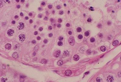
精巣18.精娘細胞と精子細胞.
ヒト、ブアン液で固定、パラフィン包埋、H-E染色、x 750.
「精巣17」の拡大。画面の中央やや右に1個のセルトリー細胞の核があり、-----
その右に接して4個の精娘細胞が認められ、これらがセルトリー細胞の細胞体
に付着している状態も明らかである。基底膜に接している円形の核は精祖細胞
の核であり、画面の左端で基底膜から離れて高い位置にある3個の大きな核は
精母細胞の核である。これら以外の管腔の近くにある小型の核は全て精子細胞
の核である。画面の中央下部で基底膜の外に付着している細胞はライディッヒ
の間細胞である。--------------------------------------------------------------------------
Testis
18. Secondary spermatocytes and spermatids.
Human, fixation with Bouin’s fluid, embedding with paraffin, H-E stain, x 750.
Higher magnification of “Testis 17”. In the middle locates one Sertoli cell and right----
to
it 4 secondary spermatocytes are recognized, embraced by the cytoplasm of this-----
Sertoli cell. Attaching to the basement membrane numerous sprematogonia are seen.
At left end of this field 3 large round nuclei of the primary spermatocytes are noted---
upper to a nucleus of Sertoli cell. Other than these, all small round nuclei are that of--
the spermatids. At lower middle an interstitial cell of Leydig attaches to the outside-----
of the basement membrane.--------------------------------------------------------------------------