精巣29.精管2.
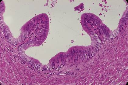
ヒト、フォルマリン固定、パラフィン包埋、H-E染色、x 200.
精管の上皮は、始めは精巣上体管の上皮と同じ2列の円柱上皮で、小さい
円形の核を持つ基底細胞と、丈の高い円柱細胞とからなる。後者は自由表
面に不動毛をそなえているが、遠位に行くにつれて不動毛は丈が低く、不
著明になる。基底細胞も次第に少なくなって、消失し、結局、上皮は単層
円柱上皮となる。少量の結合組織(粘膜固有層)に裏打ちされた上皮は、
縦走するヒダとなって管腔内に突出する。このヒダは遠位に行くにつれて
数も多く、突出の程度も高度になり、遠位端の精管膨大部では突出したヒ
ダが分岐と吻合を繰り返して、管腔は迷路のようになる。-------------------
Testis
29. Ductus deferens 2.
Human, fixation with formalin, embedding with paraffin, H-E stain, x 200.
The
epithelium of the ductus deferens is at first the same as that of the-------
ductus
epididymidis,i.e. pseudostratified columnar epithelium consisting-------
of tall columnar cells with stereocilia and small basal cells. During the course
to the distal end, the basal cells become less numerous and disappear; the----
stereocilia
also become less distinct. Finally the epithelium becomes simple---
columnar. The epithelium with underlining connective tissue protrudes into-
the lumen to form numerous longitudinal folds causing the highly irregular-
outline of the lumen seen in cross section.--------------------------------------------