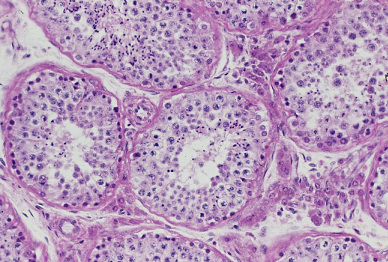
精巣3.曲精細管と間細胞.
ヒト、ブアン液で固定、パラフィン包埋、H-E染色、x 200.
中央の曲精細管の右上、右、及び左下に多数の間細胞が存在し、この曲精細管の
左上及び左下方に小動脈の横断面が見られる。この図の中の曲精細管に於いては------
精子形成の段階は様々であるが、この拡大ではその詳細を識別することはできない。
Testis
3. Seminiferous tubule and cells of Leydig.
Human, fixation with Bouin’s fluid, embedding with paraffin, H-E stain, x 200.
We can see here in the seminiferous tubules the various stages of spermatogenesis-
but no precise recognition can be done. In the loose connective tissue around the ----
tubule at center numerous interstitial cells of Leydig are visible.-------------------------