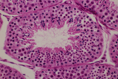
精巣8. 曲精細管2.
サル、ブアン液で固定、パラフィン包埋、H-E染色、x 300.
これは組織の固定がよくできたサルの標本である(「精巣7」と比較せよ)。基底膜に密着する
1列の細胞が精祖細胞であり、これから管腔に向かって精子に分化していく細胞が配列する。
先ず精子細胞が通常の有糸分裂で精母細胞(第一次精母細胞)を作り、これを管腔に向かって
押し上げる。精母細胞ゆっくり肥大して大きくなり、2回連続した分裂を繰り返すが、第一回
目の分裂で2個の精娘細胞(第二次精母細胞)となり、第二回目の分裂で個々の精娘細胞が2
個の精子細胞となる。結局1個の精母細胞から4個の精子細胞が生じる。4個の精子細胞はそ
れぞれ染色体数が1/2になっているので、この2回連続の分裂を成熟分裂または減数分裂とい
う。以上の経過から分かるように、精母細胞と精娘細胞、精娘細胞と精子細胞が同一の区画内
に共存することはない。図の中央を占める曲精細管では、左下方に大きな核をもった4個の精
母細胞があり、その右側に続く下壁の小さな核をもった細胞群は精子細胞である。精母細胞の
左上から上壁にかけて存在する、前2者の核の中間の大きさの核をもった細胞群が精娘細胞で
ある。これらの細胞群に接して管腔内に見られる赤く濃染した細胞体をもつ小型の細胞は精子
組織形成の途中の精子細胞である。このように精祖細胞から始まって、精母細胞・精娘細胞・
精子細胞と全部が揃って観察できる断面は、サルにおいても稀にしか見られない。―――――――――
Testis
8. Tubulus seminiferus contortus 2.
Monkey, fixation with Bouin’s fluid, embedding with paraffin, H-E stain, x 300.
This is a well preserved monkey testis (Compare with “Testis 7”! ). The spermatogenic---------- --
cells
are concentrically arranged from the basement membrane to the lumen. ----------------------
The
starting cells, the spermatogonia, constitute the outermost layer closely
attaching------------
to the basement membrane. The primary spermatocytes, composing the next layer, perform-----
the meiosis; by the first meiotic division each of them divides into two secondary spermatocytes,
which in turn by the second meiotic division, each into two spermatids. Thus the primary—―----
spermatocytes do not coexist with the secondary spermatocytes, and also the secondary----------
spermatocytes do not coexist with spermatids in the same component. In this tubule, at lower--
left
corner there are 4 large nuclei of the primary spermatocytes, and right to them
there are--
a lot of spermatids. The secondary spermatocytes occupy the upper half of this tubule. The-―---
innermost layer directly surrounding the whole lumen is constituted by the spermatids--------―-
performing the spermatohistogenesis.-------------------------------------------------------------------------