 肝臓 22.ヘリング管 1.
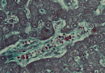
ヒト、ブアン液で固定、パラフィン包埋、M-G染色、x 160.
これは小葉間結合組織の末端部で、その中を1本の小葉間静脈の
縦断面が貫いている。この小葉間静脈右端に接して1個のヘ-----
リング管の横断面が認められる。これは4個の核が極めて狭い----
管腔を囲んでいる管である。この小葉間結合組織を囲む肝実質は-
肝細胞索と類洞からなり、肝細胞索にはいたるところに胆毛細管--
が認められる。緑色に染まっているのは結合組織繊維(細網繊維と
膠原繊維)である。----------------------------------------
Liver 22. Canal of Hering 1.
Human, fixation with Bouin's fluid, embedding with paraffin, M-G
stain, x 160.
In this figure the terminal portion of interlobular connective
----
tissue locates in the middle obliquely and there an interlobular--
vein runs longitudinally. At the right end of this vein transverse
-
section of one canal of Hering is seen, consisting of 4 small
round
nuclei. Hepatic parenchyma surrounding this connective tissue
--
consists of hepatic cell cords, in which numerous bile canaliculi
are
well observed, and sinusoids. Connective tissue fibers(reticular
--
and collagenous fibers)stains green.
------------------------
戻る